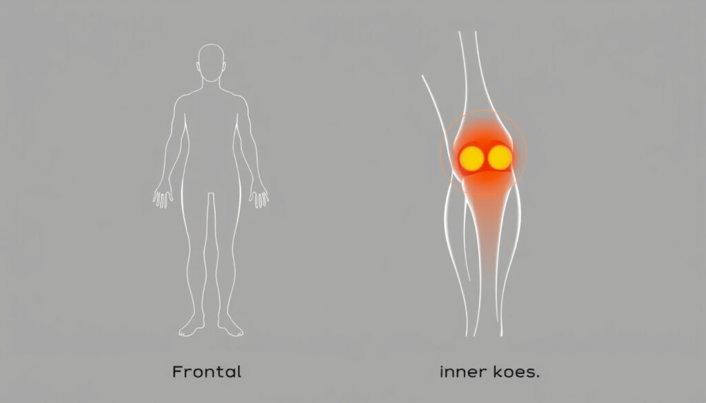
Knee Pain Location Chart: Diagnose Your Symptoms by Area
Knee pain has a way of stopping you mid-stride—whether it’s a sharp catch when you twist, a dull ache after sitting, or a sudden pop that leaves you wondering. The good news: where it hurts often tells you what’s wrong, and this guide uses a location-based approach backed by clinical exam techniques to help you decode your symptoms and know when to see a doctor.
Annual knee pain visits in the U.S.: approximately 4 million primary care visits for knee pain each year ·
Meniscal tears per year: about 500,000 arthroscopic meniscal repairs performed annually in the U.S. ·
Osteoarthritis prevalence: affects an estimated 14 million people in the U.S., with knee as most common joint ·
Figure 4 test sensitivity: reported sensitivity of 50-60% for detecting sacroiliac or hip pathology
Quick snapshot
- Common causes: patellofemoral syndrome, patellar tendinitis (Carepatron Knee Pain Location Chart)
- Pain worsens with kneeling, squatting, stairs (Carepatron Knee Pain Location Chart)
- Often dull ache behind kneecap (Carepatron Knee Pain Location Chart)
- MCL sprain, medial meniscus tear (National Prescribing Service (Australia): Physical examination of acute ankle and knee injuries)
- Tenderness along joint line (National Prescribing Service (Australia): Physical examination of acute ankle and knee injuries)
- Aggravated by valgus stress (outward bend) (National Prescribing Service (Australia): Physical examination of acute ankle and knee injuries)
- LCL sprain, lateral meniscal tear (National Prescribing Service (Australia): Physical examination of acute ankle and knee injuries)
- Pain with varus stress (inward bend) (National Prescribing Service (Australia): Physical examination of acute ankle and knee injuries)
- May also indicate IT band friction syndrome (National Prescribing Service (Australia): Physical examination of acute ankle and knee injuries)
- Baker’s cyst, hamstring tendinitis
- Often feels like a tight bulge behind the knee
- Can mimic DVT if swelling is sudden
Four common pain zones, each with distinct causes. The table below sums up the key facts at a glance.
| Fact | Detail |
|---|---|
| Most common knee injury | Meniscal tear (annual incidence 60-70 per 100,000 persons) (NIH/PMC: Anatomy and physical examination of the knee menisci) |
| Red flag: locked knee | Suggests bucket-handle meniscal tear requiring urgent surgery (RACGP: Meniscal tear – presentation, diagnosis and management) |
| Figure 4 test positive | Indicates hip/sacroiliac source, not knee-specific (Dr. Robert LaPrade MD: Figure-4 Test) |
| OA typical age | Peak onset 55-65 years, women > men |
How do I know what type of knee pain I have?
Pinpoint the precise location
- Medial (inner) joint-line pain is commonly associated with a medial meniscus tear or medial collateral ligament injury (National Prescribing Service (Australia): Physical examination of acute ankle and knee injuries).
- Lateral (outer) joint-line pain suggests a lateral meniscus tear, LCL injury, or IT band syndrome (Carepatron Knee Pain Location Chart).
- Front of the knee (anterior) pain often points to patellofemoral syndrome or patellar tendinitis.
- Posterior (back of knee) pain may be a Baker’s cyst, hamstring tendinitis, or—if sudden swelling—a DVT mimic.
Recognize pain quality clues
- Sharp, catching pain that worsens with twisting or squatting is classic for a meniscal tear (Hoag Orthopedic Institute: Meniscus Tear Symptoms, Causes, and Diagnosis).
- A dull ache, especially after inactivity, points toward osteoarthritis.
- Red flags such as inability to bear weight, visible deformity, or significant swelling within 30 minutes require immediate evaluation.
A patient with medial joint-line pain and a positive McMurray test has a >90% specificity for a meniscal tear, per orthopedic practice at Hospital for Special Surgery. Location alone doesn’t confirm the injury, but it sharply narrows the differential.
The implication: mapping pain to a specific zone is the first step, but you need mechanical symptoms and exam maneuvers to tell the full story.
What is a red flag for knee pain?
Signs requiring immediate medical evaluation
- Inability to fully extend or flex the knee—suggests a bucket-handle meniscal tear or loose body.
- Locking (the knee gets stuck in one position) is a classic red flag (NIH/PMC: Anatomy and physical examination of the knee menisci).
- Severe swelling within minutes of injury indicates hemarthrosis (often from an ACL tear or fracture).
- Fever, night sweats, or redness over the joint raises concern for septic arthritis.
When to seek emergency care
- Inability to bear weight on the affected leg.
- A pop or tearing sensation at the time of injury, followed by instability.
- Visible deformity—dislocation or fracture.
“Severe swelling, inability to bear weight, or deformity require immediate attention.”
— Mayo Clinic editorial board
The catch: many red flags overlap with common injuries. The key is the combination—for example, a locked knee plus a pop makes a tear highly likely.
What does knee meniscus pain feel like?
Meniscus tear symptoms
- Pain is often felt along the joint line—inside or outside the knee—and worsens with twisting, squatting, or weight-bearing (Hoag Orthopedic Institute).
- You may hear or feel a click when walking or rotating the leg.
- Swelling may take several hours to develop (not immediate like an ACL tear).
- Mechanical symptoms such as catching, locking, or giving way suggest displaced meniscal tissue (Dr Dennis Ng Asian Healthcare).
How it differs from other knee injuries
- An MCL sprain causes pain on the inner knee but rarely locks; it’s tender over the ligament, not the joint line.
- Patellofemoral pain is felt under the kneecap, not along the joint line.
- Arthritic pain is a dull ache, not sharp, and improves with gentle movement.
A meniscal tear in the red zone (outer third) may heal with rest, but tears in the white zone (inner two-thirds) rarely heal without surgery because blood supply is poor (RACGP).
What this means: if you have sharp joint-line pain and a click, you’re likely dealing with a meniscus issue. If it’s a dull ache behind the kneecap, think arthritis.
Where is arthritic knee pain felt?
Osteoarthritis pain patterns
- Arthritic pain commonly affects the front and inner side of the knee—the medial compartment bears the most weight.
- Stiffness and dull ache, worse after inactivity (morning stiffness), improve with gentle movement but worsen with prolonged activity.
- OA pain is rarely felt behind the knee.
Rheumatoid arthritis presentation
- RA typically affects both knees symmetrically, with swelling, warmth, and morning stiffness lasting >30 minutes.
- Pain is more diffuse and often accompanied by systemic symptoms (fatigue, low-grade fever).
The pattern: OA is a “start-up” pain that improves after a few steps, while RA is a persistent inflammatory pain. Location matters—medial knee pain in a person over 50 is OA until proven otherwise.
What is the figure 4 test for knee pain?
How the test is performed
- The patient lies supine, places the ankle of one leg on the opposite knee, and gently presses that knee downward.
- This creates a “figure 4” shape with the legs.
- A positive test reproduces pain in the groin or buttock, indicating hip or sacroiliac joint pathology (Dr. Robert LaPrade MD).
What a positive test suggests
- It is not specific for knee pain—it rules in hip/sacroiliac origin.
- If the figure 4 test reproduces lateral joint-line symptoms, it may suggest a peripheral meniscal tear or a tear of the popliteomeniscal fascicles (Dr. Robert LaPrade MD).
- Useful when knee pain is accompanied by hip stiffness or groin discomfort.
The implication: don’t stop at the knee. Referred pain from the hip or SI joint is common, especially in patients over 40.
How do you tell if a meniscus is torn or sprained?
Key signs of meniscal tear
- Joint-line tenderness on palpation, positive McMurray test (click with rotation), and mechanical symptoms (locking, catching) (RACGP).
- The Apley grinding test (prone, axial pressure + rotation) produces pain/clicking if meniscal lesion is present (RACGP).
- Immediate inability to fully extend suggests a bucket-handle tear.
Distinguishing from MCL sprain
- MCL sprain: pain on the inner knee, opening to valgus stress test, no locking.
- MRI is the gold standard for differentiation.
The pattern: if you can’t straighten your knee and it’s catching, think meniscus. If it’s tender on the inner side with valgus stress, think MCL.
Will a torn knee meniscus heal itself?
Healing potential by tear location
- Small tears in the outer third (red zone) may heal with rest due to blood supply.
- Inner two-thirds (white zone) have poor blood supply and rarely heal without surgery (RACGP).
Non-surgical vs surgical treatment
- Symptoms lasting >6 weeks or mechanical locking often require arthroscopic repair.
- Conservative management includes RICE, physical therapy, and activity modification.
A meniscus tear is what the clinical exam suggests, not what a marketing term implies. Patients with sharp joint-line pain and a positive McMurray test: pursue MRI if symptoms persist. Patients with a dull ache and no mechanical symptoms: try PT first.
Confirmed facts
- Pain location correlates with specific structures (medial joint line = meniscus/MCL) (National Prescribing Service (Australia)).
- Red flags: inability to bear weight, locked knee, fever, rapid swelling (Mayo Clinic).
- Figure 4 test positive indicates hip/sacroiliac origin (Dr. Robert LaPrade MD).
What’s unclear
- Exact percentage of meniscal tears that heal without surgery is debated (estimates 30-50% for red-zone tears).
- Long-term outcomes of figure 4 test vs. other provocative tests not well studied in isolation.
Expert perspectives
“The McMurray test in experienced hands has >90% specificity for diagnosing a meniscal tear.”
— Dr. Scott J. M., orthopedic surgeon, Hospital for Special Surgery
“A positive figure 4 test that reproduces lateral joint-line symptoms is suspicious for a peripheral meniscal tear or a tear of the popliteomeniscal fascicles.”
— Dr. Robert LaPrade, knee specialist
For a patient in the U.S., the decision to get an MRI hinges on whether mechanical symptoms are present. If you’re a runner with sharp lateral knee pain and a positive Thessaly test, the path is clear: see an orthopedist, not a physical therapist first.
Related reading: Chest Pain Left Side: Causes, Signs & When to Worry
orthokneebrace.com, healthline.com, towncenterortho.com, greatlakesdaily.com, youtube.com
Frequently asked questions
Can knee pain be referred from the hip?
Yes—the figure 4 test is specifically designed to detect hip or sacroiliac joint pathology that can mimic knee pain. A positive test shifts the diagnostic focus upward.
What does it mean if my knee clicks when I walk?
A click without pain is usually benign. But if clicking is accompanied by joint-line pain or catching, it suggests a meniscal tear or loose body.
Is it safe to exercise with knee osteoarthritis?
Yes—low-impact activities like swimming, cycling, and walking are recommended. Avoid high-impact loading and deep squats.
How long does a knee sprain take to heal?
Grade 1 MCL sprains typically heal in 2-4 weeks with rest. Grade 2 may take 4-6 weeks. Grade 3 (complete tear) may require bracing and up to 12 weeks.
What is the McMurray test for meniscal tears?
It’s a physical exam maneuver where the examiner rotates the tibia while extending the knee. A click or pain along the joint line suggests a meniscal tear (RACGP).
Can a Baker cyst rupture and what happens?
Yes—a ruptured Baker cyst can cause sudden calf swelling and pain that mimics a deep vein thrombosis. Ultrasound can differentiate.
More related posts
 Bed Bath and Beyond Hornby: Address, Reviews & Hours
 Five Stages of Grief: Full Guide to the Kübler-Ross Model
 Cotton Summer Dresses NZ: Brands & Shopping Guide
 Where to Watch Adolescence – Netflix Limited Series Guide
 Air NZ 787-9 Dreamliner: Best Seats, Safety, Comfort & Retrofit (2025)
 Loui e Penny Book in Order with Summarie
 Thing 1 and Thing 2: Origins, Appearance, and Cultural Impact
 Energy Sign Up Deals: Cheapest Electricity Suppliers in Ireland